


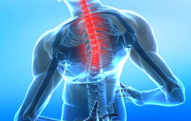
Osteochondrosis of the spine is a complex of dystrophic and degenerative changes in the intervertebral discs and the surrounding surfaces of the vertebral bodies, which are associated with the destruction of tissues and disruption of their structure. Osteochondrosis of the cervix, chest and lumbar region can be distinguished according to the level of injury.
Symptoms
The main signs that can indicate the presence of cervical osteochondrosis are a local change in the configuration of one of the segments of the cervix (development of lordosis, kyphosis or scoliosis) - a distinct visual curvature of the spine. In the longitudinal or transverse plane. The second most common symptom is pain syndrome, which can be localized not only in the area of the spine, but also in areas innervated by the corresponding nerve root in the body. Another complaint of these patients is a feeling of discomfort and fatigue in the neck.
Pain in cervical osteochondrosis usually manifests itself in the neck area and can be given to the shoulders and scapula, it can be confused with pain during a myocardial infarction since it has similar symptoms. Also, cervical osteochondrosis can be accompanied by frequent headaches, dizziness. Compression (suppression) of the arteries supplying the brain can reveal signs of brain dysfunction (neurological symptoms): nausea, vomiting, tinnitus, mood swings, anxiety, and more.
According to the severity of the pain, they are divided into 3 degrees:
- The pain occurs only with pronounced movements of the spine;
- The pain is relieved by a certain position of the spine;
- The pain is constant.
Forms
Among the syndromes caused by osteochondrosis are:
- Compression syndromes - arise by compression (radiculopathy - compression of nerve roots, myelopathy - contraction of muscles, neurovascular - compression of blood vessels and nerves);
- Reflex (muscular-tonic, neurodystrophic, neurovascular);
- Myoadaptive Syndrome (Excessive loading of healthy muscles when they are impaired by damaged muscle functions).
ᲛReasons
The mechanism of disease development is damage to the intervertebral disc for various reasons and its displacement with loss of spinal damping (relieving pressure) functions. The immediate cause of disc damage may be age-related degenerative changes associated with disruption of the blood supply to the intervertebral discs, mechanical damage to the joints, and strenuous physical exertion on the spinal column — for example, overweight.
An active role in the development of osteochondrosis is played by a sedentary lifestyle, during which a disruption of the blood supply and the functioning of the intervertebral joints develop. The mechanism of development of the disease is as follows: If the fibrous ring connecting the vertebral bodies is damaged, the intervertebral disc moves back and forth - in the lumen of the spinal canal, or laterally - forming the median and lateral disc. Hernias. The disc can enter the body of the spine itself to form an umbilical hernia - microscopic rupture of the cartilaginous tissue of the intervertebral disc into the spongy tissue of the spine. If the disc is moved backwards, it is possible to compress the spinal cord and the roots extending from it, developing a typical pain syndrome.
Diagnosis
The diagnosis of osteochondrosis of the spine is made on the basis of complaints, medical history, clinical examination and instrumental examination methods. Diagnostic measures are to clarify the causes that led to the development of neurological symptoms.
The anamnesis can be used to determine the presence of trauma, the nature of the work - constant physical overload (weight lifting), poor posture, work characteristics and the position of the spinal column at the table and while walking; Existence of infections.
In general clinical trials (clinical blood test, general urine analysis), biochemical blood analysis has no independent significance. They are prescribed to assess the current condition, to diagnose the underlying disease and developing complications.
Diagnosis is based on the clinical picture of the disease and is carried out by the method of sequential exclusion of similar diseases with clinical signs. The most common and available of the instrumental diagnostic methods is radiological examination (spondylography is a non-contrast study). It reflects the narrowing of the interstitial space and allows you to identify osteophytes (bone formations) on the vertebral bodies, but provides only indirect information about the degree of damage to the intervertebral discs.
Accurate diagnosis can be made by CT and MRI (computed tomography and magnetic resonance imaging) diagnostics, even in the early stages of the disease. CT allows you to determine the minimum abnormalities in bone and cartilage tissues, MRI - to perform a detailed analysis of soft tissue structures and determine the localization of the disc herniation.
Duplex ultrasound scan of the cerebral arteries is performed in case of suspected blood supply to the brain.
Differential diagnosis is made with diseases that have similar clinical manifestations: pathologies manifested by radiating pain in the shoulder and scapula (diseases of the liver, gallbladder, pancreatitis - inflammation of the pancreas); Cervical lymphadenitis - enlargement of the cervical lymph nodes, rheumatoid arthritis; Oncological diseases (tumors of the spine, roots, spinal cord and membranes), tumors of the pharynx and pharyngeal space, pancreatic cancer (compression of the shoulder girdle in lung cancer), metastases in the cervix; Tuberculous spondylitis - an inflammatory disease of the spine caused by Mycobacterium tuberculosis; Arachnoid cysts; Pseudocytes of dura mater; Spinal anomalies; Fibromyalgia is a disease that causes pain in muscles, ligaments, and tendons, a syndrome of chest compression - a disorder caused by excessive pressure on a neurovascular bundle that runs between the anterior and middle scapular muscles, under the first rib, and under the collarbone. Syndrome and shoulder girdle - a chronic, pathological condition caused by the formation of local muscle spasms or rings, represented by pain points.
Basic laboratory tests used:
- Clinical blood test;
- Blood chemistry.
Basic instrumental studies used:
- Spinal radiography (spondylography);
- Magnetic resonance imaging (MRI);
- Computed tomography (CT);
- Ultrasound duplex scan of cerebral arteries (if abnormal blood supply to the brain is suspected).
Additional instrumental studies used:
- Densitometry - measurement of bone density (according to indications).
Treatment
Treatment of spinal osteochondrosis depends entirely on the stage and degree of development of osteochondrosis. At the initial stage it is possible to use preventive measures, physiotherapy exercises, exercise on the trainers and fitness. During severe pain syndrome, the patient needs physical rest. Anti-inflammatory and antispasmodics are prescribed. To open the abnormal circle, it is possible to perform paravertebral blockades with anesthetics, when the pain causes muscle spasm, and the intervertebral disc is more strongly compressed, which in turn intensifies the pain.
Warming ointments are used topically on the skin in the spine to improve local blood supply to the skin and reduce tissue swelling. These patients are shown with a corset. Chondroprotectors are effective in patients with early stages of osteochondrosis - drugs that improve cartilage repair, as well as drugs that improve the local blood supply, venotonics, B vitamins. In cases where the pain syndrome does not stop. From a medical point of view it has been a long time and there is a clinic for spinal cord root compression with intervertebral hernia, surgical removal of the damaged intervertebral disc has been shown. In case of total compression of the spinal cord by the disc, early surgery is indicated.
You do not have to wait until a person starts urinating or defecating spontaneously - in which case the spinal cord injury may already be irreversible. As physiotherapy procedures, magnetotherapy, ultrasound, massage, manual therapy, acupuncture and physiotherapy exercises are prescribed.
Complications
Possible vegetative-vascular dystonia and cardiac dysfunction, cerebrovascular accident, hypotension and hypertension (decrease and increase in blood pressure), vestibular disorders (movement coordination disorder), vertebral artery syndrome (disease caused by obstruction of the spine)) Shoulder joint.
Prevention
For the prevention of osteochondrosis it is necessary to deal with its causative factors, namely: to prevent spinal injuries, to fight stress (increase in severity) on the spine with excess weight. For those who are already suffering from the initial stage of osteochondrosis, it is recommended to wear a corset at home and during physical activity. In order for the spine to relax during sleep, it is recommended to sleep on an orthopedic mattress and pillows.
What questions to ask your doctor
Are there exercises to relieve symptoms?
What medications help to fight cervical osteochondrosis?
What if you do not start treatment on time?
Patient advice
In exercise, weight loss in the presence of excess weight, the use of cool or warm compresses helps to alleviate the symptoms of osteochondrosis of the thoracic spine. Proper nutrition, spine monitoring, treatment of chronic diseases, and injury prevention are also important.